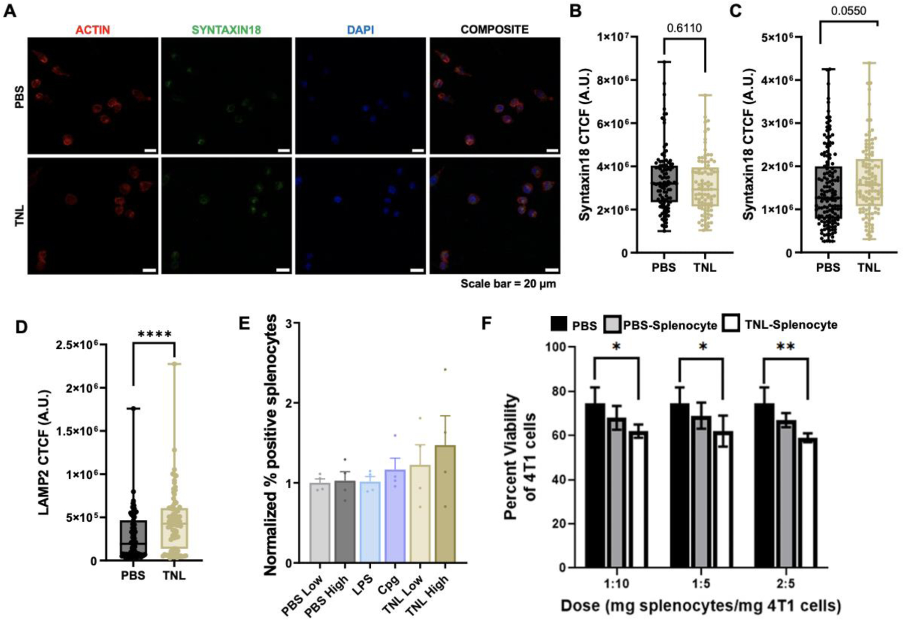
Figure 3. DC phagocytosis and functional analyses.

(A) Syntaxin18 24 h representative micrograph. Syntaxin18 expression after (B) 4 h and (C) 24 h. (D) LAMP2 expression after 24 h. (E) CD3+ Tetramer staining. (F) Functional viability assay treating 4T1 breast cancer cells with TNL-treated splenocytes. Scale bar = 50 μm. *p<0.05, **p<0.01, ****p<0.001.
