FIG. 1.
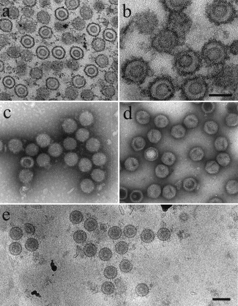
Electron microscopy of HSV-1 procapsids. (a) Thin section showing procapsids in the nucleus of a BHK cell infected for 15 h with HSV-1 mutant m100; (b) thin section preparation of m100 procapsids after isolation by antibody precipitation; (c) m100 procapsids after negative staining with uranyl acetate; (d) negatively stained HSV-1 B-capsids; (e) m100 procapsids preserved in the frozen hydrated state. Note that m100 procapsids are round in profile (c and e) and consist of distinct shell and core layers. All micrographs are shown at the same magnification (bar = 1,500 Å) except for panel b (bar = 1,000 Å).

