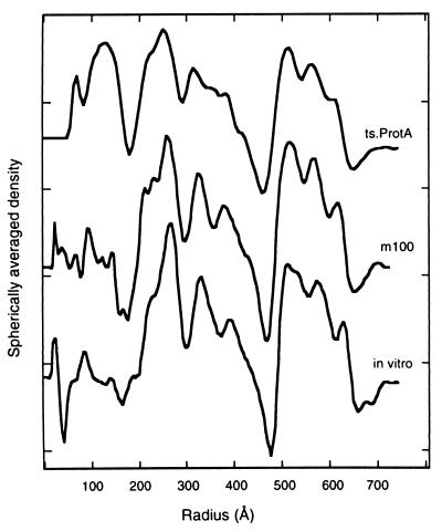
FIG. 5.
Radial density profiles of HSV-1 procapsids from three sources: native procapsids isolated from BHK cells infected at the NPT with tsProt.A (top curve), native procapsids isolated from BHK cells infected with m100 (middle), and procapsids assembled in vitro from lysates of Sf9 cells containing HSV-1 capsid proteins (bottom) (46). The profiles were calculated by spherical averaging of the corresponding three-dimensional density maps. The main features of the surface shell (three peaks between radii of 480 and 650 Å) and the scaffold (three peaks between radii of 200 and 460 Å) are well conserved. However, the tsProt.A procapsids are the only ones to show a significant density peak inside the scaffold shell suggested to correspond to the HSV-1 UL26 gene product, the virus protease.

