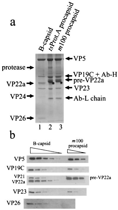
FIG. 7.
Protein composition of HSV-1 procapsids. SDS-polyacrylamide gel electrophoresis of m100 and tsProt.A procapsids was followed by staining with Coomassie blue (a) and Western immunoblotting with specific antibodies (b). Similar analyses of HSV-1 B-capsids are shown for comparison. The starred bands in lanes 2 and 3 of panel a were identified as actin as described in the text. The positions of the HSV-1 protease (UL26 gene product) and the antibody heavy and light chains (Ab-H and Ab-L) are indicated in panel a. Specimens in panel b were loaded for electrophoresis in twofold dilutions. The amount of sample loaded in the VP26 row was twofold greater than that loaded in other rows. Note that procapsid bands identified as VP5, VP19C, pre-VP22a, and VP23 were stained with specific antibodies, and VP26 was not detected in either m100 or tsProt.A procapsids.

