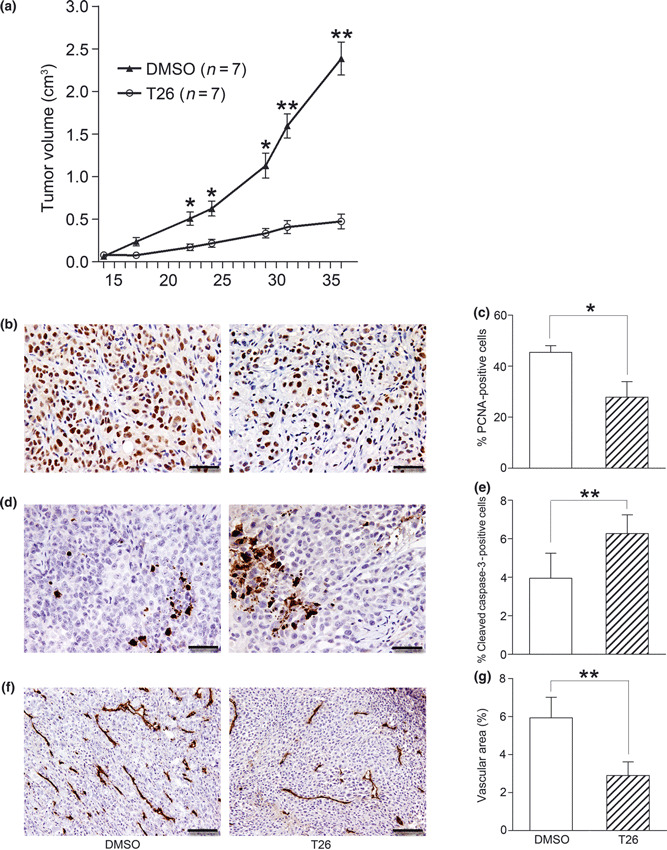
Figure 7.

Effects of T26 on in vivo tumor growth. Fourteen days after nude mice had been injected with 2 × 10⁶ PCI66 cells, T26 treatment was initiated as described in the Materials and Methods. (a) Tumor sizes were determined twice a week and are shown here as the mean ± SEM (n = 7). *P < 0.05, **P < 0.01 compared with 0.1% dimethyl sulfoxide (DMSO) control. (b–g) Tumor tissues obtained 36 days after injection of tumor cells were subjected to immunohistochemical analysis using (b,c) anti‐proliferating cell nuclear antigen (PCNA), (d,e) anti‐cleaved caspase‐3, and (f,g) anti‐CD31 antibody. (b,d,f) Representative results from three mice are shown. Bars, 50 μm (b,d); 100 μm (f). (c,e,g) The number of PCNA‐ (c) or cleaved caspase‐3‐positive cells (e) was determined and CD31‐positive areas (g) were measured in five randomly chosen visual fields at a magnification of ×100 using ImageJ software (National Institutes of Health, Bethesda, MD, USA). Data are shown as the mean ± SD (n = 4). *P < 0.05, **P < 0.01.
