
Figure 1.
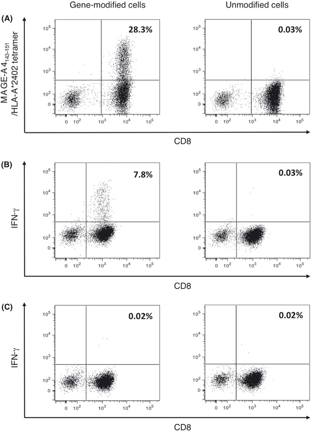
Transduction of melanoma‐associated antigen (MAGE)‐A4‐specific T‐cell receptor (TCR) in human lymphocytes. Peripheral blood mononuclear cells from healthy donors were stimulated with anti‐CD3 mAb and interleukin‐2. Cells were cultured with or without retroviral vector encoding MAGE‐A4‐specific TCR, designated gene‐modified or unmodified cells, respectively. (A) Representative staining for gene‐modified and unmodified cells with MAGE‐A4143–151/HLA‐A*2402 tetramer and antihuman CD8 mAb are shown. (B,C) Gene‐modified and unmodified cells were stimulated with T2‐A*2402 cells pulsed with the MAGE‐A4143–151 peptide (B) or HLA‐A*2402‐binding irrelevant control peptide (C). Representative specific intracellular interferon (IFN)‐γ staining is displayed. Numerical value indicates the percentage of the tetramer+ cells or IFN‐g+ cells among CD8+ cells.