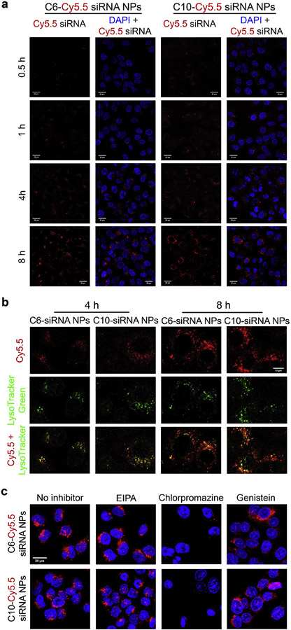
Figure 4.
Cellular uptake of C6-Cy5.5-siRNA NPs and C10-Cy5.5-siRNA NPs. a) Internalization kinetics of NPs within 0.5-8 h. The NPs were prepared following the procedure in in vitro delivery assays. After incubation with NPs for 0.5-8 h, A549 cells were washed with PBS, fixed by 4% PFA, and stained with DAPI for confocal microscopy. Scale bars = 20 μm. b) Co-localization (yellow) of siRNA-Cy5.5 (red) and lysozymes (green) at 4 and 8 h. The cells were incubated with LysoTracker green for 0.5 h before fixing and staining. Scale bars = 10 μm. c) Cellular uptake in the presence of various endocytosis inhibitors (8 h). Scale bars = 20 μm.

