. 2024 Jun 6;15(1):2360601. doi: 10.1080/19491034.2024.2360601

Figure 2.

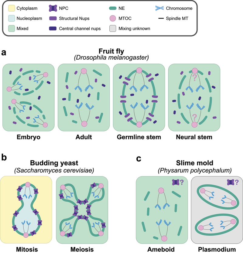
Part a. Illustrated representations of the types of nuclear division found in the fruit fly Drosophila melanogaster. Multinucleated embryos undergo semi-open divisions with local NEBD at the poles of the cell, full NPC disassembly, and extranuclear centrosomes, the MTOCs in flies. Adult somatic cells undergo complete NEBD with full NPC disassembly and extranuclear centrosomes. Germline stem cells undergo semi-closed divisions, with central channel nucleoporins disassociating from the core nucleoporins and centrosomes embedded in the NE. Neural stem cells undergo semi-closed divisions with full NPC disassembly, but the positioning of the centrosomes is unknown. Part b. Illustrated representations of the types of nuclear division found in the budding yeast Saccharomyces cerevisiae. The yeast undergoes closed mitotic division, but a semi-closed division during meiosis. The central nucleoporins are not dissociated from the core nucleoporins in either division. Part c. Illustrated representations of the types of nuclear division found in the slime mold Physarum polycephalum. Cells undergo open divisions during their amoeboid life cycle stage but switch to divisions with intact NEs in the multinucleated plasmodium stage. It is not known if there is nucleocytoplasmic mixing in plasmodium, and the state of the NPCs is not known for either stage.

Distinct nuclear division strategies are used in different developmental contexts within the same organism. a. Cartoon representing the four currently identified types of nuclear division found in the fruit fly Drosophila melanogaster. From left to right: partial nuclear envelope breakdown (NEBD) in the embryo [20], open division in adult somatic cells, semi-closed division with partial NPC disassembly in female germline stem cells [18], and semi-closed division with complete NPC disassembly in neural stem cells [19]. The position of the MTOC (centrosome) in fly neural stem cells is currently unknown, represented with question marks. b. Cartoon representations of the budding yeast Saccharomyces cerevisiae nucleus undergoing closed mitosis (left) [52]; and semi-closed meiosis (right) [17,53]. c. Cartoon representations of the slime mold Physarum polycephalum nucleus undergoing an open division in the cell’s ameboid stage of development (left) and a closed division during the syncytial plasmodium form (right) [54]. It is currently unknown if the nuclear pore complexes (NPCs) disassemble in either mode of division, represented with question marks. Nuclear lamina is omitted for simplicity. MTOC (Microtubule Organizing Center).