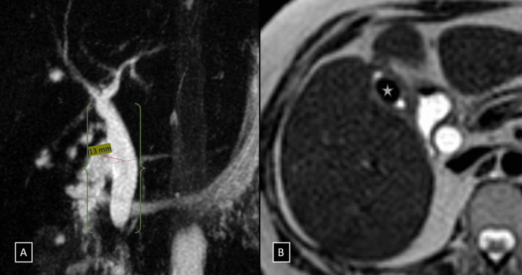
Figure 1. (A and B) Coronal MIP images from heavily T2-weighted pulse sequences showing a dilated CBD with abrupt tapering just proximal to the ampulla of Vater (denoted by a green brace in figure A). Axial sections of T2-weighted imaging showing a large ovoid calculus measuring 13 × 9 mm within the lumen of the gallbladder (white star in B).
CBD, common bile duct

