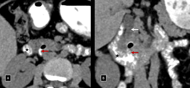
Figure 2. (A and B) Axial and coronal reformatted images of NCCT with positive oral contrast administration demonstrating a small periampullary diverticulum (red arrows) with internal air foci arising from the medial wall of the distal second part/proximal third part of the duodenum (black star), compressing the retro-pancreatic portion of the distal CBD extrinsically (white arrow), just proximal to the ampulla of Vater.
NCCT, non-contrast enhanced computed tomography; CBD, common bile duct

