. 2024 Jun 10;13:RP93094. doi: 10.7554/eLife.93094

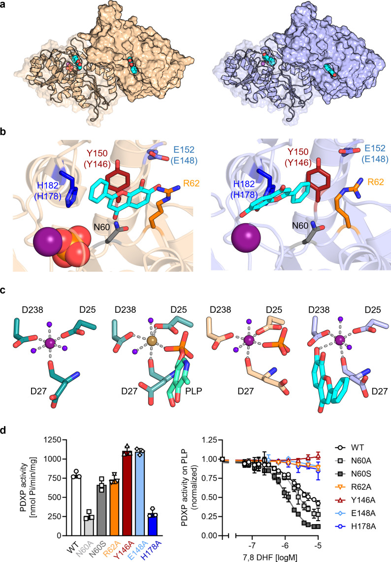
Figure 3. X-ray crystal structures of human pyridoxal 5’-phosphate phosphatase (PDXP) in complex with 7,8-dihydroxyflavone (7,8-DHF).

(a) The models were refined to a resolution of 1.5 Å for full-length human 7,8-DHF-PDXP with phosphate (PDB code 9EM1, colored in wheat yellow, left panel) and 1.5 Å for full-length human 7,8-DHF-PDXP without phosphate (PDB code 8S8A, colored in light blue, right panel). One protomer of each homodimeric PDXP is shown in cartoon representation and the other protomer in surface representation. 7,8-DHF is displayed in sphere representation with its C-atoms in cyan. Mg2+ ions are shown as deep purple spheres and phosphate ions are shown in sphere representation with the phosphorous atom in orange. (b) Orientation of 7,8-DHF in the active sites of human 7,8-DHF-PDXP in the presence or absence of phosphate. Structural details of bound 7,8-DHF and adjacent residues of the active sites are shown. Left, phosphate-containing 7,8-DHF-PDXP (wheat yellow, cartoon representation). Right, phosphate-free 7,8-DHF-PDXP (light blue, cartoon representation). 7,8-DHF is shown in stick representation (cyan C-atoms). The corresponding amino acids in murine PDXP are given in parentheses (see also Figure 3—figure supplement 1e and f). (c) Comparison of the Mg2+ coordination spheres. From left to right: human apo-PDXP (PDB: 2P27), human PDXP in complex with pyridoxal 5’-phosphate (PLP) (PDB: 2CFT), human PDXP in complex with 7,8-DHF in the presence of phosphate (PDB: 9EM1), human PDXP in complex with 7,8-DHF in the absence of phosphate (PDB: 8S8A). The catalytically essential Mg2+ is shown as a deep purple sphere. In 2CFT, Mg2+ was exchanged for Ca2+, which is shown here as a light brown-colored sphere. Water molecules are shown as blue spheres. (d) Verification of 7,8-DHF-PDXP interactions. Left panel, phosphatase activity of purified PDXP or the indicated PDXP variants. Data are mean values ± SD of n=3 biologically independent experiments. Right panel, determination of the IC50 values of 7,8-DHF for purified PDXP or the indicated PDXP variants. Data are mean values ± SD of n=3 biologically independent experiments. Apparently missing error bars are hidden by the symbols.

Figure 3—source data 1. Phosphatase activity and 7,8-dihydroxyflavone (7,8-DHF) sensitivity of pyridoxal 5’-phosphate phosphatase (PDXP) and PDXP variants (to Figure 3g).
Data are OD values and normalized data of malachite green assays, using pyridoxal 5’-phosphate (PLP) as a PDXP substrate.

Figure 3.

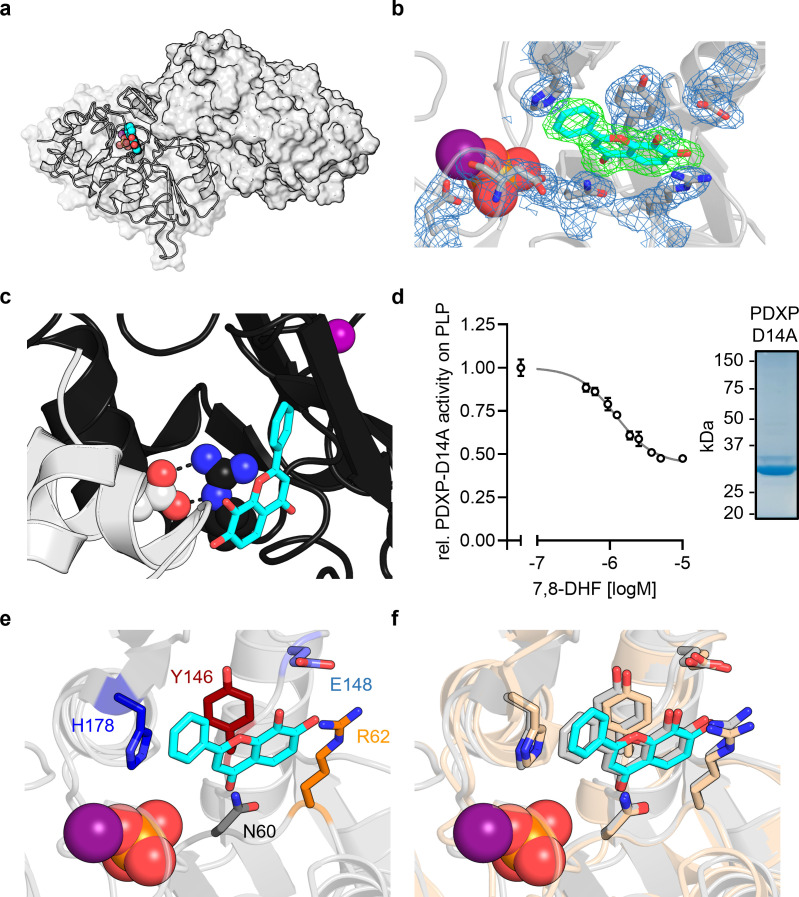
Figure 3—figure supplement 1. X-ray crystal structures of murine pyridoxal 5’-phosphate phosphatase (PDXP) in complex with 7,8-dihydroxyflavone (7,8-DHF).

Figure 3—figure supplement 1.

(a) X-ray crystal structure of the assembled homodimeric, full-length murine PDXP in gray cartoon (protomer A) and surface (protomer B) representation. The model was refined to a resolution of 2.0 Å (PDB code 8QFW). 7,8-DHF is shown in sphere representation with its C-atoms in cyan. The Mg2+ ion is shown as deep purple sphere. The phosphate ion is shown as spheres with the phosphorous atom in orange. (b) The 2Fo Fc electron density map of the depicted amino acids is contoured at a root mean square deviation (RMSD) of 1.0 in blue mesh and superimposed with the refined model. The Fo Fc polder electron density map of 7,8-DHF is contoured at an RMSD of 3.0 in green mesh. (c) A salt bridge between Arg62 in the B-protomer (in black) and Asp14 of a symmetry-related A-protomer (in gray) blocks the 7,8-DHF binding site in the crystal lattice of murine PDXP. 7,8-DHF (in stick representation with cyan C-atoms) is modeled based on the A-protomer. (d) In vitro phosphatase activity of the purified PDXP-D14A variant in the presence of 7,8-DHF. Data are mean values ± SD of n=3 technically independent experiments. Apparently missing error bars are hidden by the symbols. The purity of PDXP-D14A is shown in the Coomassie blue-stained gel on the right. (e) Structural details of bound 7,8-DHF and adjacent residues of the active site of mPDXP (in gray cartoon representation). (f) Superimposition of the 7,8-DHF binding sites in the phosphate-containing murine PDXP (shown in gray) and human PDXP (in wheat yellow) structures. The corresponding amino acids are shown as gray or wheat yellow-colored sticks. 7,8-DHF bound to murine or human PDXP is shown as sticks colored in gray or cyan, respectively. The position of the Mg2+ and phosphate ions is based on human PDXP.
Figure 3—figure supplement 1—source data 1. Effect of 7,8-dihydroxyflavone (7,8-DHF) on the phosphatase activity of pyridoxal 5’-phosphate phosphatase (PDXP)-D14A.
Data are OD values and normalized data of malachite green assays, using pyridoxal 5’-phosphate (PLP) as a PDXP substrate.
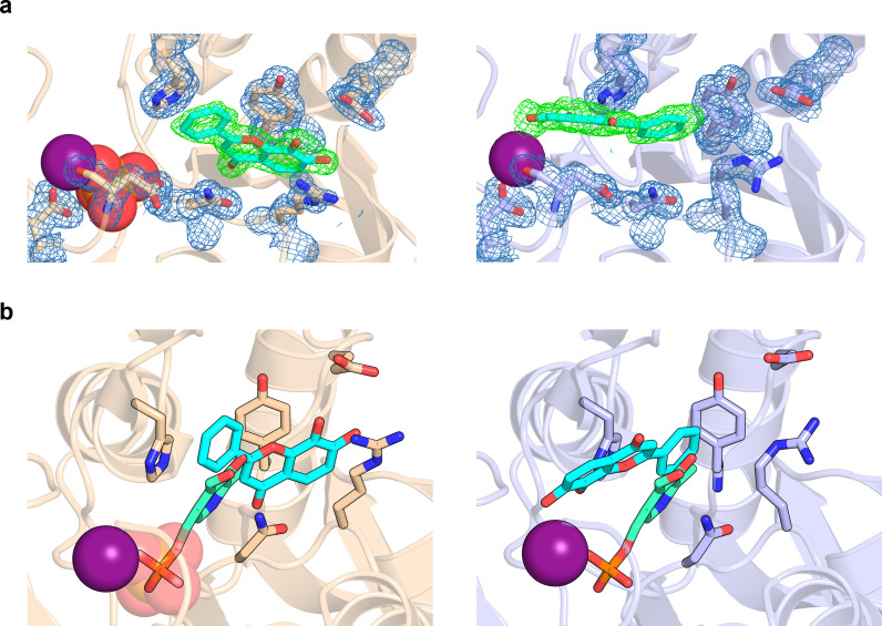
Figure 3—figure supplement 2. 7,8-Dihydroxyflavone (7,8-DHF) coordination in pyridoxal 5’-phosphate phosphatase (PDXP).

Figure 3—figure supplement 2.

(a) The 2Fo Fc electron density map of the depicted amino acids is contoured at a root mean square deviation (RMSD) of 1.0 in blue mesh and superimposed with the refined model. The Fo Fc polder electron density map of 7,8-DHF is contoured at an RMSD of 3.0 in green mesh. The Mg2+ ion is shown as a deep purple sphere. The phosphate ion is shown as a sphere with the phosphorous atom in orange. Left panel, human PDXP with phosphate (cartoon representation in wheat yellow); right panel, human PDXP without phosphate (cartoon representation in light blue). (b) Comparison of the 7,8-DHF and pyridoxal 5’-phosphate (PLP) binding sites in human PDXP with phosphate (wheat yellow, left panel) or human PDXP without phosphate (light blue, right panel). 7,8-DHF is shown in stick representation with cyan C-atoms. PLP (in stick representation with green C-atoms) was modeled based on a superposition of the human PDXP-PLP complex (PDB code 2CFT).
Figure 3—figure supplement 3. Alignment of human and murine pyridoxal 5’-phosphate phosphatase (PDXP).
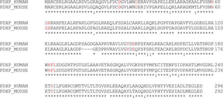
Figure 3—figure supplement 3.

Protein sequences of human PDXP (UniProtKB Q96GD0) and murine PDXP (UniProtKB P60487) were aligned with the EMBL-EBI multiple sequence alignment tool Clustal Omega version 1.2.4. PDXP residues found to engage in 7,8-dihydroxyflavone (7,8-DHF) interactions (highlighted in red color) are identical in human and murine PDXP.
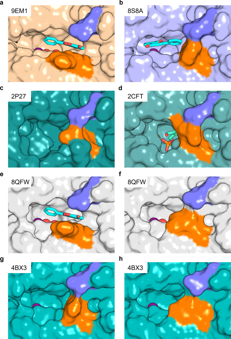
Figure 3—figure supplement 4. Salt bridge formation between Glu152 (Glu148) and Arg62 gates the active site entrance in pyridoxal 5’-phosphate phosphatase (PDXP).

Figure 3—figure supplement 4.

Shown are views of the active site entrance in (a) hPDXP + 7,8-DHF with PO43-, (b) hPDXP + 7,8-DHF without PO43-, (c) apo-hPDXP, (d) hPDXP + pyridoxal 5’-phosphate (PLP), (e) mPDXP + 7,8-DHF with PO43, chain A, (f) mPDXP + 7,8-DHF with PO43-, chain B (inhibitor-free), (g) apo-mPDXP, chain A, (h) apo-mPDXP, chain B. The cap domain residue Glu152 in hPDXP (corresponding to Glu148 in mPDXP) is shown in blue, and the core domain residue Arg62 in hPDXP and mPDXP is shown in orange. 7,8-Dihydroxyflavone (7,8-DHF) is shown in stick representation with C-atoms in cyan. PLP is shown in stick representation with C-atoms in light green. The phosphate is shown in sphere representation with the phosphorous atom in orange. Mg2+ is shown as a deep purple sphere. hPDXP, human PDXP; mPDXP, murine PDXP. The respective PDB entries are indicated.
Figure 3—figure supplement 5. Purity of the employed pyridoxal 5’-phosphate phosphatase (PDXP) and PDXP variants.
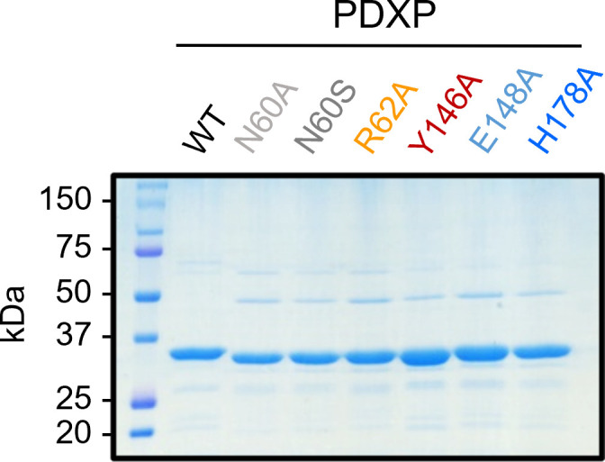
Figure 3—figure supplement 5.

A Coomassie blue-stained gel is shown. Source data are available for this figure.