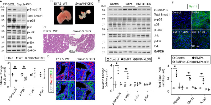
(A) Activation of the intracellular downstream Smad1, p38, Jnk, and Erk signaling pathways in wildtype (WT) and Bmpr1a conditional knockout (CKO) lung tissues was detected by the western blot (WB) and quantified by densitometry. The levels of protein phosphorylation were normalized by the corresponding total protein and is presented as a relative change to the WT, *p<0.05. (B) Gross view of whole lungs from Smad1/5 double conditional knockout mice (Smad1/5 CKO) and WT littermates showed that simultaneous deletion of Smad1 and Smad5 in lung mesenchyme completely disrupted lung development. (C) No airway dilation or cysts were observed in the hematoxylin and eosin (H&E)-stained tissue sections of the Smad1/5 CKO lungs at embryonic day (E)17.5. (D) Expression of airway smooth muscle cells (SMCs) and elastin was not altered in E17.5 Smad1/5 CKO lungs, as shown by immunostaining of Cdh1, Acta2, and elastin. (E) Changes of intracellular signaling pathways in cultured fetal lung mesenchymal cells upon treatment with BMP4 (50 ng/ml) and/or LDN193189 (200 nM) were detected by WB and quantified by densitometry. The relative change to the control condition is presented, *p<0.05. (F and G) Altered expression of SMC genes at the protein level (Myh11) and the mRNA level (Myocd, Myh11, and Acta2) was respectively analyzed by immunostaining and real-time PCR for the primary culture of E15.5 WT lung mesenchymal cells treated with BMP4 (50 ng/ml), LDN (200 nM), and SB (1 µM), *p<0.05.
Figure 4—source data 1. Original file for the western blot (WB) analysis in Figure 4A (anti-p-Smad1/5).
Figure 4—source data 2. Original file for the western blot (WB) analysis in Figure 4A (anti-total Smad1, anti-Jnk, and anti-Erk).
Figure 4—source data 3. Original file for the western blot (WB) analysis in Figure 4A (anti-p-p38).
Figure 4—source data 4. Original file for the western blot (WB) analysis in Figure 4A (anti-p38).
Figure 4—source data 5. Original file for the western blot (WB) analysis in Figure 4A (anti-p-Jnk).
Figure 4—source data 6. Original file for the western blot (WB) analysis in Figure 4A (anti-p-Erk).
Figure 4—source data 7. Original file for the western blot (WB) analysis in Figure 4A (anti-GAPDH).
Figure 4—source data 8. PDF containing Figure 4A and original scans of the relevant western blot (WB) analysis (anti-p-Smad1/5, anti-total Smad1/5, anti-p-p38, anti-p38, anti-p-Jnk, anti-Jnk, anti-p-Erk, anti-Erk, and anti-GAPDH) with highlighted bands and sample labels.
Figure 4—source data 9. Original file for the western blot (WB) analysis in Figure 4E (anti-p-Smad1/5).
Figure 4—source data 10. Original file for the western blot (WB) analysis in Figure 4E (anti-total Smad1).
Figure 4—source data 11. Original file for the western blot (WB) analysis in Figure 4E (anti-p-p38).
Figure 4—source data 12. Original file for the western blot (WB) analysis in Figure 4E (anti-p38).
Figure 4—source data 13. Original file for the western blot (WB) analysis in Figure 4E (anti-p-Jnk).
Figure 4—source data 14. Original file for the western blot (WB) analysis in Figure 4E (anti-Jnk).
Figure 4—source data 15. Original file for the western blot (WB) analysis in Figure 4E (anti-p-Erk).
Figure 4—source data 16. Original file for the western blot (WB) analysis in Figure 4E (anti-Erk).
Figure 4—source data 17. Original file for the western blot (WB) analysis in Figure 4E (anti-GAPDH).
Figure 4—source data 18. PDF containing Figure 4E and original scans of the relevant western blot (WB) analysis (anti-p-Smad1/5, anti-total Smad1/5, anti-p-p38, anti-p38, anti-p-Jnk, anti-Jnk, anti-p-Erk, anti-Erk, and anti-GAPDH) with highlighted bands and sample labels.

