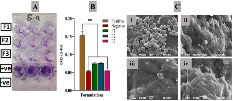
Figure 6.
Effect of various test samples, including drug-loaded nanofibers on the growth of biofilm; (A) photograph representing biofilm growth in 96-well plate and (B) plot representing a comparative reduction in biofilm after treating with nanofiber formulations. (C) SEM images depicting biofilm growth and reduction upon nanofiber treatment of Bacterial film (positive control) (i), Reduction in biofilm after treatment with nanofiber of PVP-RSV (ii), PVA-AMP (iii), and PVP-PVA-RSV-AMP (iv). All the data given as mean±S.D. **p<0.01, one-way ANOVA followed by Bonferroni multiple comparison test. All the data represented as mean±SD (n=3).

