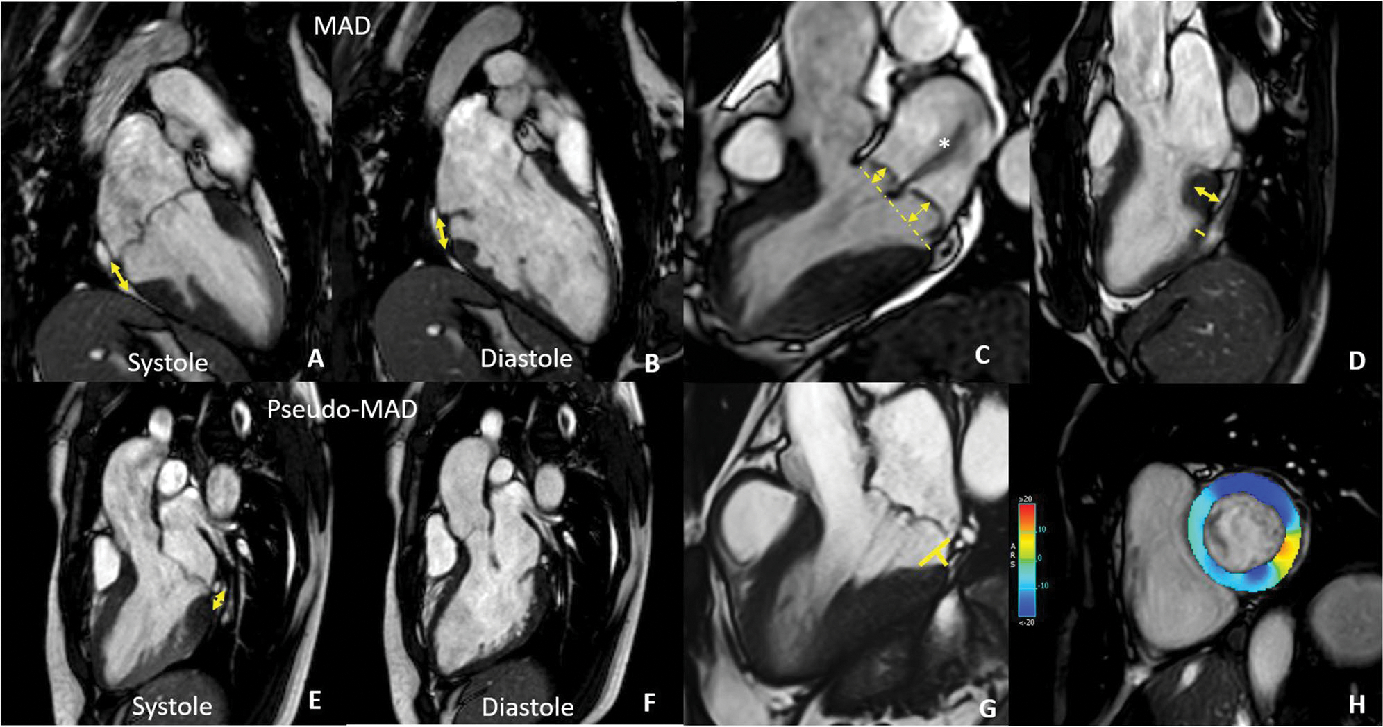
Fig. 3.

CMR findings in MVP. In A and B are reported systolic (A) and diastolic (B) frames of the same patient showing mitral annulus disjunction (double-headed arrow) that needs to be distinguished from pseudo-MAD (E and F) due to the juxtaposition of the posterior leaflet on the atrial wall in systole. C 3-chamber long axis showing severe bileaflet mitral valve prolapse with high prolapse volume and a huge jet of regurgitation (asterisks). D Basal LV hypertrophy with a ratio of LV thickness between basal and mid segments of the inferolateral wall > 1.5 at end-diastole. G Curling distance by tracing a line between the top of the LV I wall and the LA–MV leaflet junction, and from this line, a perpendicular line to the lower limit of the mitral annulus at end-systole. H GLS analysis showing contractility alteration of the inferolateral wall
