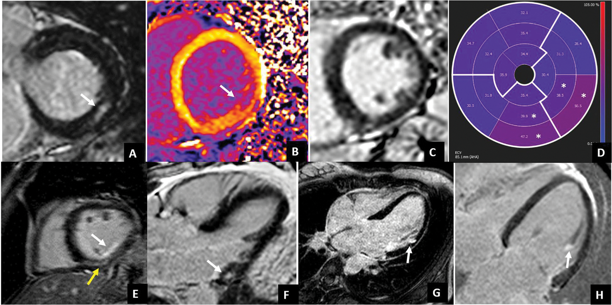
Fig. 4.

LGE patterns typically associated with arrhythmic MVP. LGE usually occurs at the level of the LV inferolateral wall. Different LGE patterns have been described: non-ischemic mid-wall LGE (white arrows in A and F, yellow arrow in E), subendocardial LGE (white arrows in E and G), papillary muscle LGE (with arrow in H). Interstitial fibrosis documented by native T1 mapping and ECV values has been found to be increased not only at the site of LGE (white arrow in native T1 map in B) but also in LGE negative patients (case example in C) with diffusely high ECV values which are higher in the inferior and inferolateral mid-basal wall (white asterisks)
