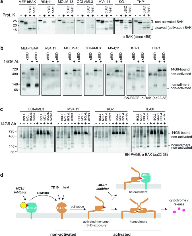
Fig. 4. BAK is not constitutively activated in blood cancer cell lines.
a The majority of BAK in untreated leukaemia cell lines is resistant to proteinase K. bak−/−bax−/− MEFs expressing hBAK and the indicated cancer cell lines were permeabilized and incubated with cBID (cBIDM97A) or incubated at 43 °C (heat) to activate BAK. Membrane fractions were incubated with proteinase K and blotted for BAK (as in Fig. 2b). Data are representative of at least two independent experiments. b The majority of BAK in untreated leukaemia cell lines is gel-shifted by 14G6. Membrane fractions from (a) were incubated with or without 14G6, run on BN-PAGE, and blotted for BAK (as in Fig. 1c). Data are representative of at least two independent experiments. c BAK activation by BH3 mimetic treatment demonstrated by loss of 14G6 binding. Four acute myeloid leukaemia cell lines were incubated with 1 µM iBCL2 (venetoclax), iMCL1 (S63845), iBCLxL (A-1331852), alone or in combination for 3 h. Membrane fractions were then incubated with or without 14G6, and analyzed as in (b). Cell lysates were also assessed for BAK and BAX cleavage by proteinase K (Fig. S6). Data are representative of at least two independent experiments. d Schematic of BAK activation by various stimuli demonstrating that in the cell types examined, the majority of BAK becomes activated only after apoptotic signalling. Here, MCL1 represents prosurvival proteins, as MCL1 inhibitor alone could trigger BAK activation in three cell lines (Figs. 4c and S6).

