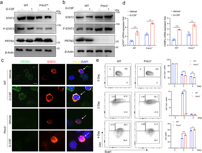
Fig. 5. Deficiency of PRTN3 promotes STAT3-dependent myeloid differentiation.
a protein expression of STAT3, P-STAT3, PRTN3, and β-Actin were analyzed in HEK293T cells with empty vector or PRTN3-HA transfection to overexpress PRTN3 and then treated with 10nM G-CSF for 24 h (n = 3); (b)protein expression of STAT3, P-STAT3, PRTN3, and β-actin were analyzed in Prtn3−/− primary Ckit+ cells with G-CSF treatment for 24 h, compared to WT cells (n = 3); c immunofluorescence staining of STAT3 in Prtn3−/− primary Ckit+ cells with G-CSF treatment for 2 h (n = 3); d messenger RNA (mRNA) of P27kpi1 and C/Ebpα were determined in Prtn3−/− primary Ckit+ cells with G-CSF treatment for 24 hours, compared to WT cells (n = 3); e Lineage- Sca-1+c-Kit+ (LSK) cells from WT and Prtn3−/− mice were used for ex vivo myeloid culture (n = 3). scale bars, 50 μm; *p < 0.05, **p < 0.01, ***p < 0.001. Data are the mean ± s.d.; n: biologically independent experiments. Statistical analysis was performed using an unpaired two-tailed Student’s t test.

