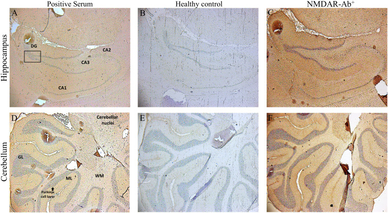
Figure 3.
Binding of sera positive for autoantibodies to subunit α4 of the α4β2-nAChRs, to rat brain tissue by Indirect Immunostaining. Sagittal whole rat brain sections were incubated with sera antibodies from the three patients with AES (A, D), from NMDAR encephalitis patients (C, F) or from healthy controls (B, E). Representative images are shown the localization of rat α4β2‐nAChRs bound by specific serum antibodies from 1 out of the 3 patients with autoantibodies against subunit α4‐nAChRs in the hippocampus (A) and the cerebellum (D), a specific positive signal which was confirmed only by the patients’ sera bearing α4-nAChR-Abs, whereas no specific binding was detected in any region of the rat brain tissue by serum components originated from 1 out of the 10 healthy controls (B, E). Staining and images of rat brain sections with serum antibodies from one NMDAR encephalitis patient as positive control (C, F) are depicted. CA1–CA3, hippocampal area cornu ammonis 1–3; DG, dentate gyrus; cerebellar layers. GCl, granular cell layer; ML, molecular layer; WM, white matter.

