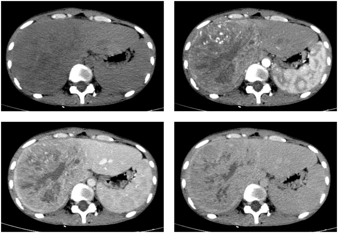
Figure 1.
CT scan images in axial views before and after the injection of a contrast agent depict a sizable liver mass with well-defined boundaries. The mass exhibits heterogeneous enhancement in both the hepatic arterial and portal venous phases, although the enhancement is less intense than the normal liver parenchyma.

