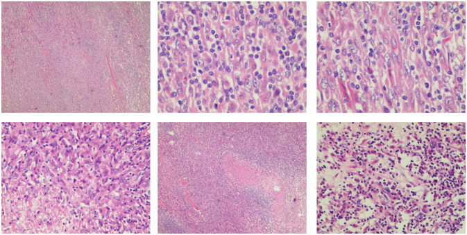
Figure 3.
Pathological image of inflammatory myofibroblasts in the liver. The tumor is composed of a proliferation of spindle cells and a large number of inflammatory cells, predominantly T lymphocytes. Collagen fiber proliferation is evident, accompanied by focal necrosis. No significant mitotic figures are observed.

