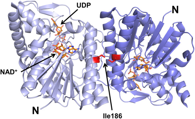
FIGURE 5.

Three dimensional structure of UXS1. Cartoon representation of the hUXS1 crystallographic dimer based on the 1.26 Å crystal structure of hUXS1 bound with NAD+ and UDP (PDB ID: 2B69). The two UXS1 monomers are shown in light and dark blue. NAD+ and UDP are shown as orange sticks. Ile186 is located in the dimer interface where it makes intermolecular hydrophobic contact with Ile186 in the other subunit.
