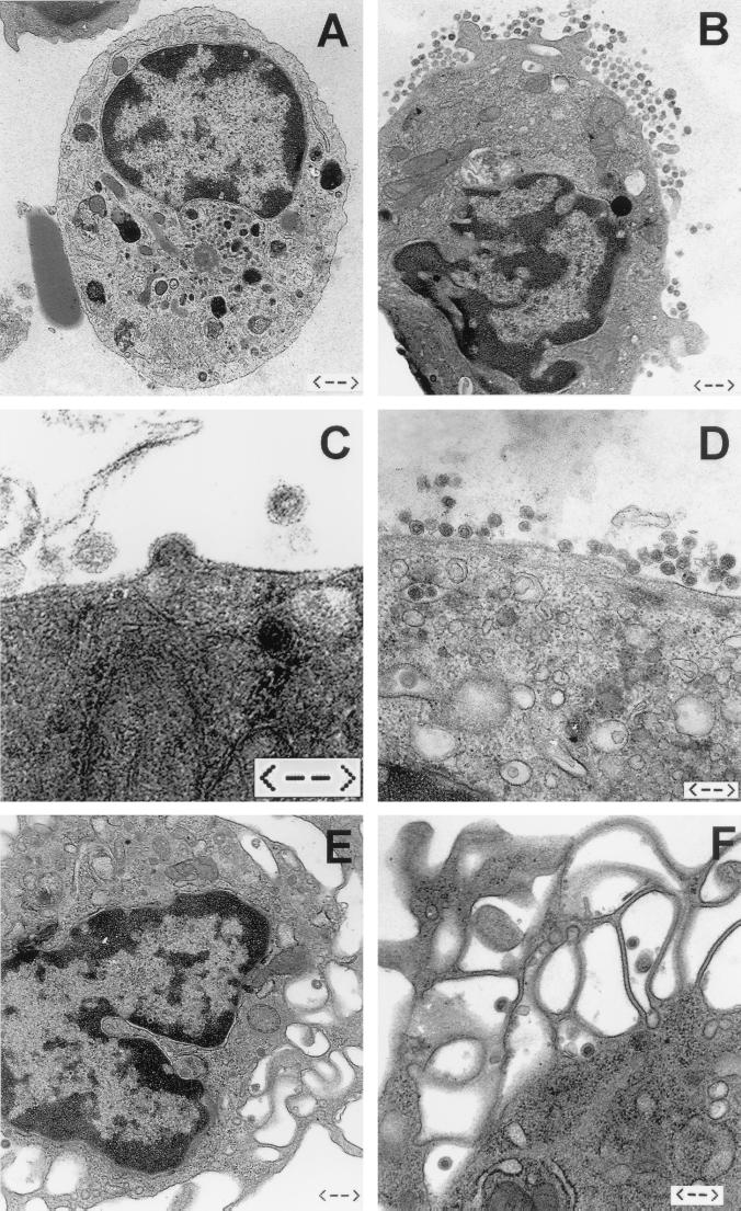
FIG. 1.
Ultrastructural analyses of microglia and macrophages isolated ex vivo. Viral particles were never found in or around microglia cells isolated from noninfected rats (A) (bar = 1.1 μm). Abundant retroviral virions were associated with microglia cells (B to D) (bars = 0.6, 0.25, and 0.4 μm, respectively) and—although fewer in numbers—with peritoneal macrophages (E and F) (bars = 0.6 and 0.4 μm, respectively) from infected rats. Budding of retroviruses from the cell membrane of microglia could be detected frequently (C), while intracellular particles were rarely found in microglia (D) and peritoneal macrophages (F).

