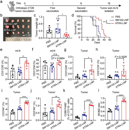
Figure 7.

a) Treatment scheme of an orthotopic CT26 colorectal tumor model using mGP70‐loaded LNPs. (b and c) Photographs and weights of isolated tumors (n = 5 biological independent samples). Statistical significance was calculated by unpaired two‐tailed Student's t‐test. d) Kaplan–Meier survival curves of mice after different treatments (n = 11–14 biologically independent samples). The percentages of CD3+CD8+ and CD3+CD4+ T cells in mLN e and f) and tumor g and h) of treated mice. The population of CD3−NK1.1+ i), CD3+NK1.1+ j), and CD86+F4/80+ cells among tumor cells. k) M1/M2 ratio in the tumor microenvironment (n = 5 biological independent samples for all flow cytometry analysis). Data are shown as mean ± SD. Statistical analysis was calculated using one‐way ANOVA and Tukey's post‐hoc test. * p < 0.05, ** p <0.01, *** p <0.001, **** p <0.0001, n.s. represents not statistically significant.
