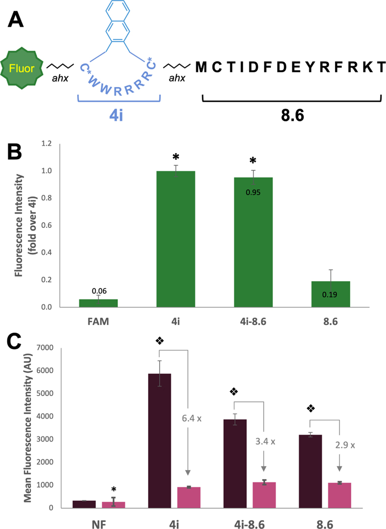
Figure 6: Peptide 4i enables delivery of cell-impermeable peptide cargo.
(A) Structure of peptide 4i-8.6. (B) Relative fluorescence intensity of FAM-labeled peptides measured by flow cytometry as compared to 4i. MDA-MB 231 cells were incubated with 5 µM FAM-labeled peptide for 2 h, trypsinized, and total cellular uptake was measured by flow cytometry (488 nm laser, 530/30A filter). Bars represent the relative geometric mean fluorescence intensity across 3 replicates. Fold change over 4i is labeled on bars. * indicates significant difference from FAM and 8.6. (C) Mean (geometric) fluorescence intensity of NF-labeled peptides measurd by flow cytometry. MDA-MB 231 cells were incubated with 5 µM NF-labeled peptides in serum-free or 10% serum media for 2 h, trypsinized, and total cellular uptake was measured flow cytometry (633 nm laser. 660/20A filter). * indicates difference from peptides in the 10% serum condition (p<0.025). Split diamond indicates significant difference from all conditions (2-way ANOVA, Holm-Sidak, p<0.003). NF is shown as an arithmetic mean as some cells exhibited negative fluorescence, preventing calculation of a geometric mean.

