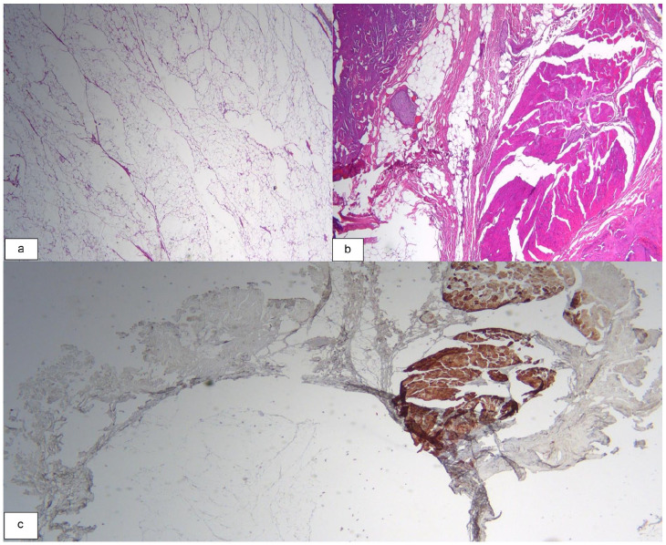
Figure 2.
(a) Histologic appearance of an overall well circumscribed intramuscular lipoma with mature univacuolated adipocytes of fairly uniform size (H&E, X20); (b) Focal area of infiltration within the surrounding skeletal muscle (H&E, X40); (c) Desmin immunostaining to highlight the skeletal muscle.

