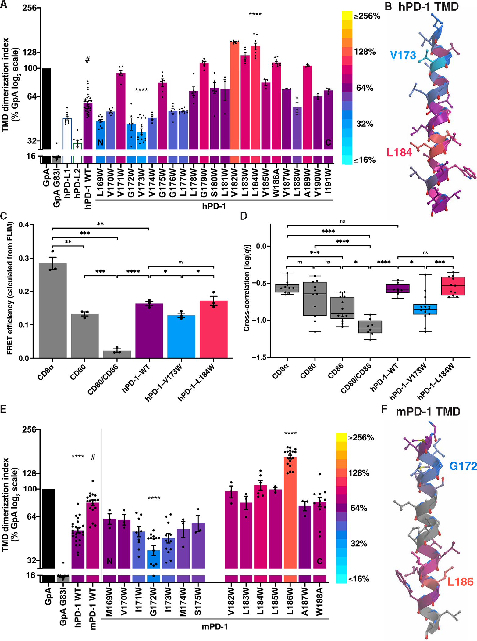
Fig. 4. hPD-1 and mPD-1 TMDs dimerize using N-terminal contacts.

(A) The effects on dimerization as determined by TOXGREEN of Trp substitutions at the indicated positions in the hPD-1 TMD. Bars were assigned a color on the basis of the log2 scaled look-up table (right). (B) Helical model of the hPD-1 TMD color-coded as in (A) with the V173hPD-1 low and L184hPD-1 high Trp substitutions labeled. (C) FRET efficiency calculated from FLIM of the indicated pairs of mEGFP- and mCherry2-tagged constructs expressed in baby hamster kidney cells. (D) FCCS measurements of mEGFP- and mCherry2-tagged constructs expressed in HEK293T cells pooled across four biological replicates. (E and F) Analysis and color-coding of the mPD-1 TMD as in (A) and (B) revealing G172mPD-1 low and L186mPD-1 high Trp substitutions. Significance in (A), (C), and (E) was determined by unpaired t tests; significance in (D) was determined by ANOVA test with Tukey’s correction for multiple comparisons: *P < 0.05, **P < 0.01, ***P < 0.001, and ****P < 0.0001.
