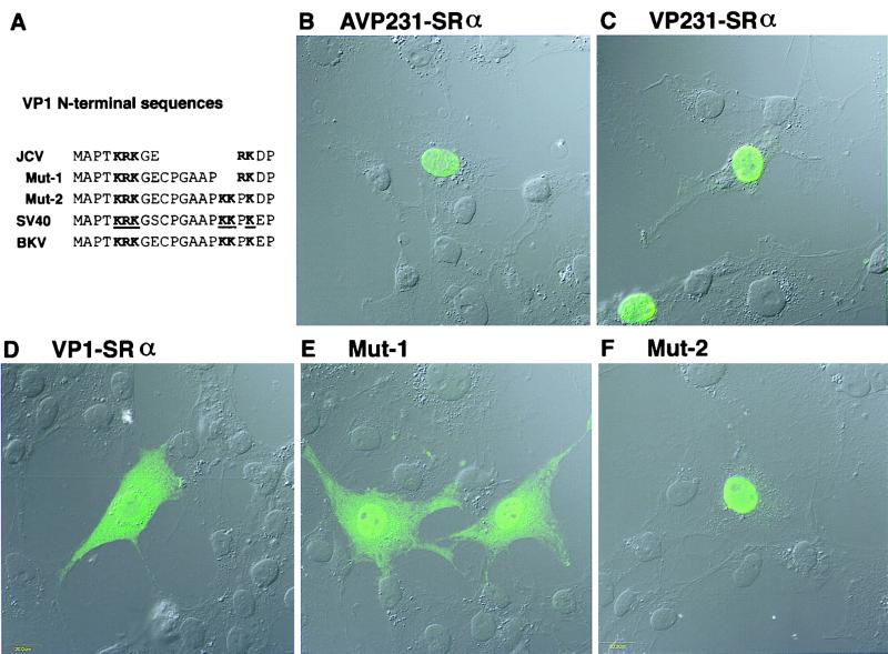
FIG. 8.
Distribution of VP1 in cells transfected with AVP231-SRα, VP231-SRα, or VP1-SRα and distribution of the two VP1 mutants, Mut-1 and Mut-2. To study nuclear transport of VP1, COS-7 cells were transfected with each of the three expression vectors, and distribution of VP1 or the VP1 mutants was investigated by immunocytochemistry using a confocal microscope. (A) The N-terminal sequences of JCV VP1, JCV VP1 mutants Mut-1 and Mut-2, SV40 VP1, and BKV VP1 are aligned. In the N-terminal sequence of JCV VP1, the basic amino acids KRK and RK (in boldface) are encoded in a monopartite structure. In contrast, in the N-terminal sequence of SV40 VP1, the two clusters of basic amino acids KRK and KKPK are encoded in a bipartite structure and identified as the NLS, as indicated with boldface and underlining (28). Mut-1 encodes the basic amino acids KRK and RK in a bipartite structure, and Mut-2 encodes KRK and KKPK in a bipartite structure. In each row of amino acid sequence, basic amino acids which are likely responsible for nuclear transport are indicated in boldface. (B and C) When cells were transfected with AVP231-SRα or VP231-SRα, VP1 was efficiently transported to the nucleus and identified as numerous speckles, indicating that VP1 is accumulated in discrete subnuclear regions, possibly with VP2 and VP3. (D) When cells were transfected with VP1-SRα, VP1 was distributed both in the cytoplasm and in the nucleus. VP1 was distributed more diffusely in the nucleus. (E) Mut-1 was transported to the nucleus more efficiently and detected more prominently in the nucleus than wild-type VP1. However, Mut-1 was still distributed both in the cytoplasm and in the nucleus. In the nucleus, Mut-1 was distributed diffusely except for nucleoli, and no speckle was identified. (F) Mut-2 was efficiently transported to the nucleus and distributed diffusely except for nucleoli. No speckle was identified in the nucleus.

