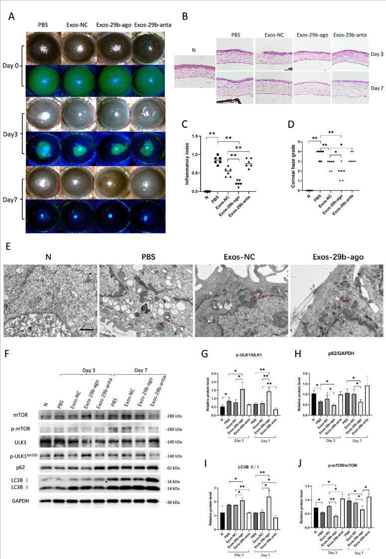
Figure 2.
Exos-29b-ago alleviated the corneal inflammatory, corneal haze , and activated autophagy in the cornea of CI mice. (A) Cornea fluorescein staining photographs and bright field photos of all groups at 3 and 7 days after treatment (day 3 and day 7). (B) H&E staining showed the histologic structure of the cornea of all groups. The eyeballs were harvested at day 3 and day 7. Scale bar = 100 µm. (C) The corneal inflammatory index of all groups at day 3 (n = 6). (D) The corneal haze grade of all groups at day 7 (n = 6). (E) TEM pictures showed there were autophagosomes (double membrane structure, red arrow) in the corneal epithelium of CI mice. Scale bar = 1.0 µm. (F) The results of expressions of the autophagy marker proteins p-mTOR, mTOR, p-ULK1Ser555, ULK1, p62, LC3BI, LC3BII, and GAPDH in CI mice of all groups at day 3 and day 7. (G) Relative expression of the ratios of p-ULK1Ser555/ULK1 (n = 3). (H) Relative expressions of p62 to GAPDH (n = 3). (I) Relative expressions of the ratios of LC3BII/I (n = 3). (J) Relative expression of the ratios of p-mTOR/mTOR (n = 3). *P < 0.05, **P < 0.01.

