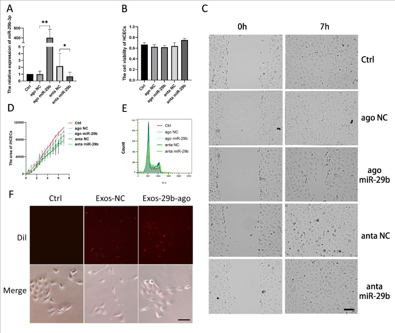
Figure 7.
Effects of miR-29b-3p in cell migration, cell proliferation and cell viability on iHCECs in vitro. (A) The results of RT-qPCR showed the levels of miR-29b-3p in iHCECs after transfection (n = 3). (B) The CCK-8 assay for cell viability of iHCECs after transfection (n = 3). *P < 0.05; **P < 0.01. (C) The results of wound healing in iHCECs showed the condition of gap at 0 hour and 7 hours, Scale bar = 50 µm. (D) The area of iHCECs within the gap (Sum Area time point- Sum Area 0 hour) of iHCECs (n = 3). (E) The results of flow cytometry for cell cycle composition of iHCECs after transfection (n = 3). (F) Dil-labeled (red) Exos-NC and Exos-29b-ago tracking in the iHCECs. Exos-NC without Dil-label set as Ctrl. The iHCECs incubated with Dil-labeled Exos for 12 hours (20 ×).

