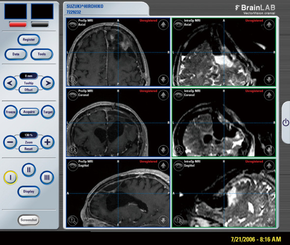
Fig. 3.

Comparison of MRI image on neuronavigation display (Left: preoperative axial, coronal, and sagittal views on T1WI. Right: intraoperative axial, coronal, and sagittal view on T2WI). Note remarkable “brain shift” after connection of ventricle with tumor removal cavity.
