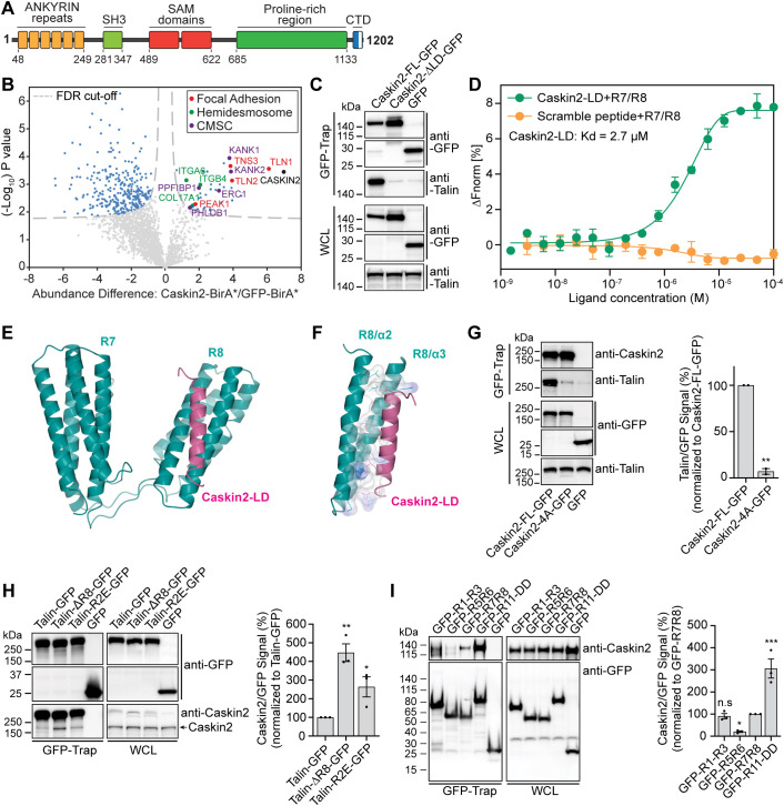
Fig. 1.
Caskin2 binds directly to talin via a C-terminal LD motif. (A) Schematic of caskin2; the C-terminal domain (CTD) contains an LD motif (white bar). (B) Proximity biotinylation assays were performed with PA-JEB/β4 keratinocytes ectopically expressing either Caskin2 fused to the biotin ligase BirA* or GFP–BirA*, as a negative control. The volcano plot shows the results from three independent experiments [threshold false discovery rate (FDR): 0.01 and S0: 0.1]. Significant proximity interactors of Caskin2 and GFP are indicated in light blue (GFP interactors, left and Caskin2 interactors, right), red (FA proteins), green (hemidesmosome proteins) or purple (CMSC components). (C) GFP-trap pulldown of Caskin2–GFP and Caskin2-ΔLD–GFP expressed in GE11tetON β1 cells, followed by western blotting for talin and Caskin2. (D) MST assay demonstrating binding of Caskin2-LD peptide to the talin R7/R8 peptide (n≥5). Peptide with scrambled amino acid sequences was used as negative control. Error bars show mean±s.d. (E) X-ray structure of the talin-R7R8 (residues 1358–1653, cyan) fragment in complex with Caskin2-LD peptide (residues 1187–1202, magenta). (F) Structure analysis show the hydrophobic interaction (with surface charge distribution) of Caskin2-LD (magenta ribbon) with talin-R8 (cyan ribbons). Residues from both talin R8 and Caskin2-LD peptide that form the interface between the two molecules are shown in sticks. The electrostatic surface of these residues are shown [blue for positive, red for negative (not present) and white for neutral]. (G) GFP-Trap pulldown of GFP, Caskin2–GFP and Caskin2-4A–GFP expressed in COS-7 cells. Proteins in the GFP-trap sample and whole-cell lysate (WCL; input) were detected by western blotting using antibodies against talin, Caskin2 and GFP. Graph shows quantification of talin binding to Caskin2-FL–GFP or Caskin2-4A–GFP from GFP-Trap pulldown experiments; n=3 independent repeats. **P<0.01 (two-tailed unpaired t-test). (H,I) GFP-Trap pulldown in COS-7 cells expressing Caskin2 (without a GFP tag) together with talin–GFP, talin-ΔR8–GFP and talin-R1523E/K1530E–GFP (H) or the indicated GFP-tagged talin polypeptides (I). Proteins in the GFP-trap sample and WCL (input) were detected by western blotting using antibodies against Caskin2 and GFP. *P<0.05, **P<0.01, ***P<0.001 (one-way ANOVA with uncorrected Fisher's LSD multiple comparisons test). Error bars in G–I are mean±s.e.m. Western blots in C, G, H and I are representative of three independent experiments.

