Fig. 5.
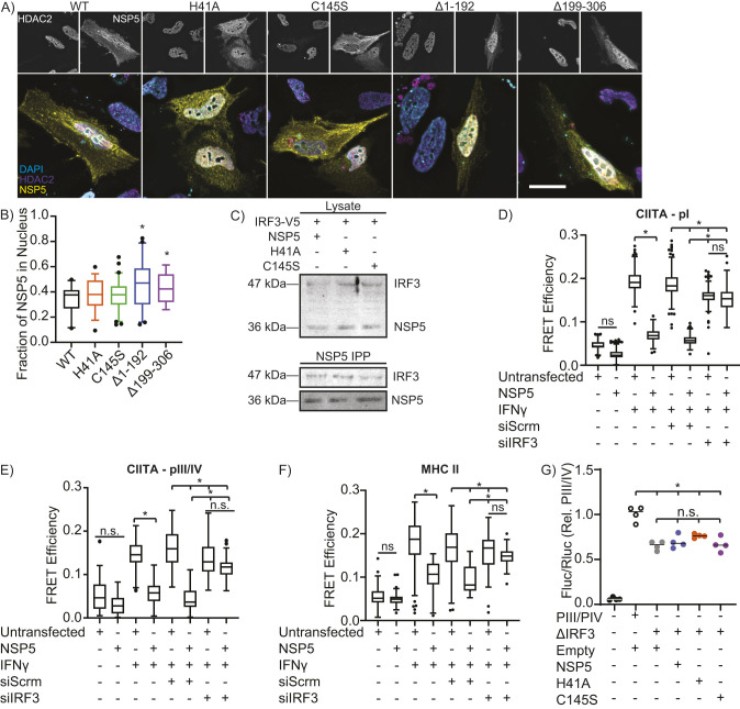
NSP5 suppresses CIITA promoter activity via IRF3. (A,B) Fluorescence micrographs (A) and quantification (B) of NSP5 (yellow) and HDAC2 (magenta) nuclear localization in HeLa cells transfected with wild-type (WT) or mutant NSP5. The nucleus has been stained with DAPI (cyan). Scale bar: 10 µm. (C) IRF3 co-immunoprecipitation with wild-type NSP5 and with the H41A and C145S NSP5 catalytic mutants. n=3. (D–F) Quantification of FISH-FRET at the CIITA pI (D), CIITA pIII/IV (E) and MHC II (F) promoters in NSP5-transfected A549 cells treated with IFNγ and a non-targeting (siScrm) or IRF3-depleating siRNA (siIRF3). (G) Impact of wild-type (NSP5) versus protease-inactivated H41A and C145S NSP5 mutants on the activity of the wild-type (pIII/IV) or IRF3-binding site deleted (ΔIRF3) CIITA pIII/IV promoter in A549 cells, as quantified by a dual-luciferase assay. Empty, cells are transfected with the empty vector used to express NSP5. Images are representative of a minimum of 30 cells imaged over three independent experiments. Data are representative of (A) or quantify (B–F) three independent experiments, and are plotted as interquartile range with median (box and line) with whiskers representing the 5–95th percentiles (B,D–F) or showing the mean (line, G). *P<0.05; n.s., not significant (Kruskal–Wallis test with Dunn correction compared to WT (B) or the indicated groups (D–G)].

