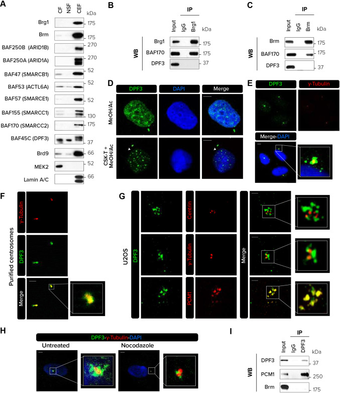
Fig. 1.
DPF3 localizes to centriolar satellites in interphase. (A) The cytosolic fraction (CF), nuclear soluble fraction (NSF) and chromatin-enriched fraction (CEF) were isolated from U2OS cells and analyzed by western blotting for the indicated proteins. (B,C) Immunoprecipitation (IP) of endogenous Brg1 (B) or Brm (C) from U2OS cells followed by western blotting (WB) for Brg1, Brm, BAF170 and DPF3. (D) U2OS cells were pre-extracted or not with CSK buffer containing Triton X-100 (CSK-T), fixed with methanol and acetone (80:20) and stained for DPF3. Individual channels and merged channels are shown. White arrowheads indicate centrosomes. (E,F) Co-staining for DPF3 (in green) and γ-tubulin (in red) in U2OS cells (E) or isolated centrosomes purified from KE37 cells (F). Individual channels, merged channels and magnifications of boxed regions are shown. (G) U2OS cells were co-stained for DPF3 (in green) and centrin, γ-tubulin or PCM1 (in red). Images were captured with Zeiss LSM 880 Airyscan high-resolution microscope. Individual channels, merged channels and magnifications of boxed regions are shown. (H) U2OS cells were treated with nocodazole (5 μg/ml) for 2 h to depolymerize the microtubule network. Cells were co-stained for DPF3 (in green) and γ-tubulin (in red). Merged channels with DAPI nuclear staining (in blue) and magnifications of boxed regions are shown. (I) Immunoprecipitation (IP) of endogenous DPF3 from U2OS cells followed by western blotting (WB) for DPF3, PCM1 and Brm. Western blotting and immunofluorescence images are representative of at least three independent experiments. Scale bars: 5 µm (D,E,H); 2 µm (F,G).

