Fig. 2.
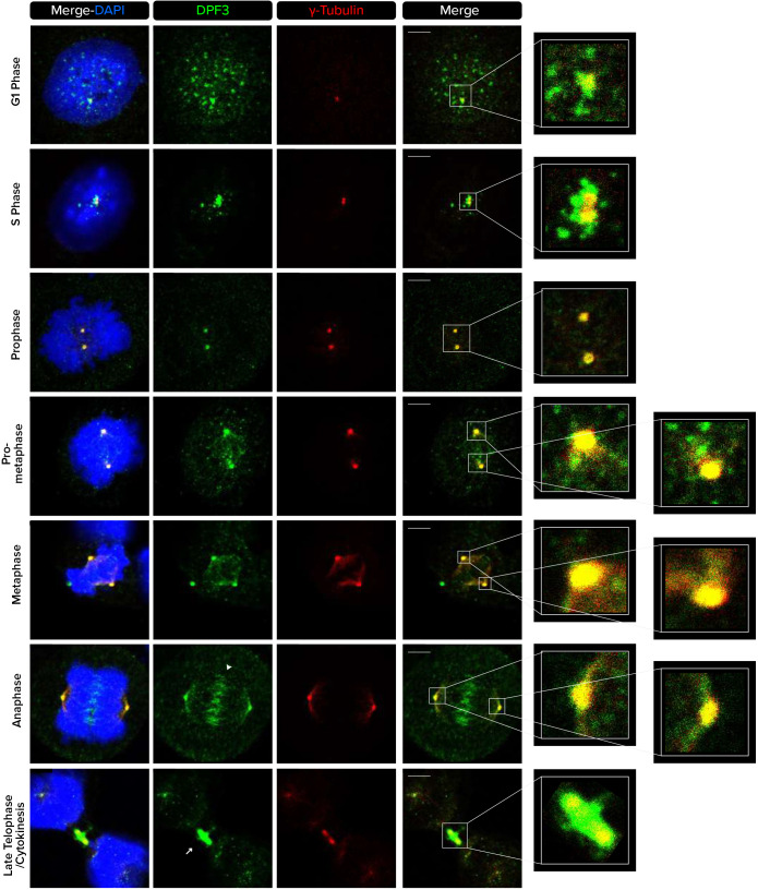
DPF3 localizes to centrosomes and microtubule-based structures during mitotic cell division. U2OS cells were co-stained for DPF3 (in green) and γ-tubulin (in red). Individual channels, merged channels with DAPI nuclear staining (in blue), and magnifications of boxed regions are shown. Nuclear condensation and the position of γ-tubulin foci identify cells in different cell cycle stages. The white arrowhead indicates the position of the spindle midzone in anaphase. The white arrow indicates the position of the midbody during cytokinesis. Images are representative of at least three independent experiments. Scale bars: 5 µm.

