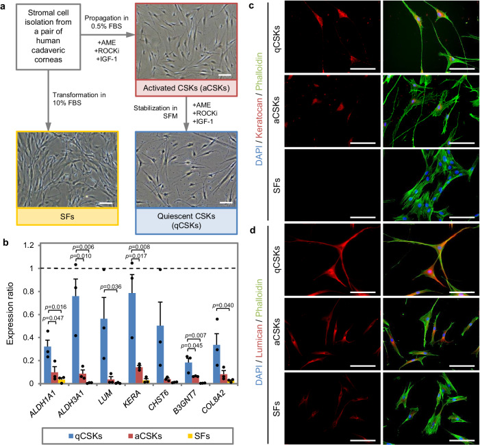
Fig. 1. Phenotypical features of cultivated corneal stromal keratocytes.
a The propagation of corneal stromal keratocytes (CSKs) was achieved by culturing the corneal stromal cells in the “activated” form in the 0.5% fetal bovine serum (FBS)-supplemented media, containing amniotic membrane extract (AME), ROCK inhibitor (ROCKi), and insulin growth factor 1 (IGF-1). The “activated” state was referred to as the activated CSKs (aCSKs). The aCSKs entered a quiescent state following culture in serum-free media (SFM), which we referred to as the quiescent CSKs (qCSKs). The qCSKs featured stellate morphology with thin cell bodies and long cell processes. As a comparison, stromal fibroblasts (SFs) that were transformed by culturing the cells in 10% serum-supplemented media had a bipolar morphology with large cell bodies and pseudopodial processes. b The qCSKs (blue bars) expressed higher levels of native CSK’s genetic markers than the aCSKs (red bars) and SFs (yellow bars). The gene expression levels were normalized to the corneal stromal tissue, indicated by the dashed line. Data were presented as mean ± SEM (n = 3 in each group). Statistical significance was assessed with one-way ANOVA, followed by post hoc Tukey test. Immunocytochemistry of keratocan (c) and lumican (d), double stained with phalloidin, confirmed the earlier morphological and gene expression analysis. The qCSKs exhibited more abundant keratocan and lumican proteins than aCSKs. In contrast, the SFs were largely absent of these proteoglycans. The staining was repeated on 3 independent cells. Scale bars = 50 µm. Source data are provided as a Source Data file.

