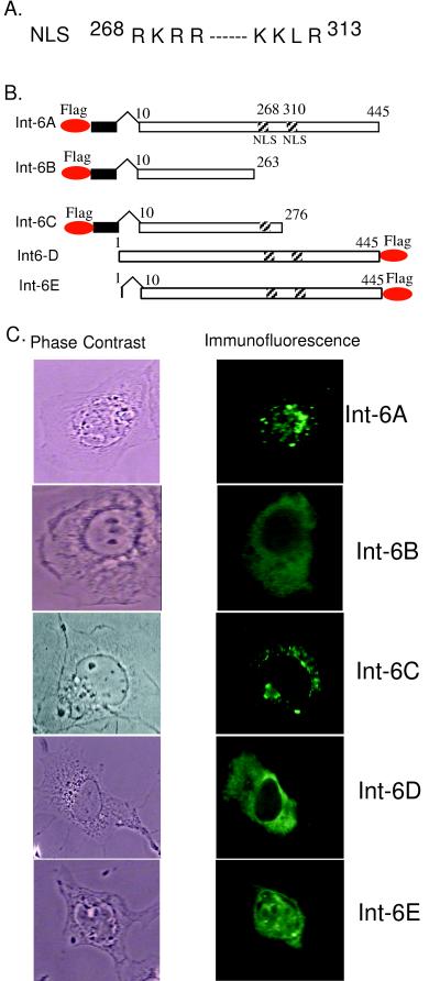
FIG. 2.
(A) Bipartite nuclear localization signal (NLS) of Int6. (B) Maps of different Int6 constructs. Oval circles represent the Flag epitope. Black rectangles represent an unrelated sequence of 13 residues. Hatched squares represent the nuclear localization signals. (C) Subcellular location of different Int6 proteins in cells. HT1080 cells was transfected with Int6A, Int6B, Int6C, Int6D, or Int6E. Twenty-four hours posttransfection, immunofluorescence was performed using the Flag probe (D-8) antibody to detect different Int6 proteins. Phase-contrast (left panel) and immunofluorescence (right panel) images are shown.

