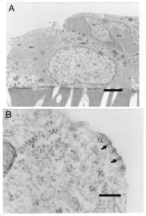
FIG. 5.
Incomplete budding at surface of MV-infected neurons. Primary CD46+ neuron cultures were infected with MV Edmonston (MOI = 1) or mock infected, fixed at 3 d.p.i. with glutaraldehyde, and processed for EM. (A) Two adjacent neuronal cell bodies containing cytoplasmic fuzzy nucleocapsids (MV) but few buds at the cell surface. Arrows indicate intact cell membranes separating the two cells. Magnification, ×7,560. Bar = 2 μm. (B) Higher magnification of infected neuron shows smooth nucleocapsid alignment at the cell surface, but only at the immature stage of budding. Magnification, ×96,600. Bar = 200 nm. Closed large arrows, cross-sectional view of nucleocapsid; open large arrow, longitudinal view of nucleocapsid; N, nucleus; M, mitochondria; R with small arrows, ribosomes.

