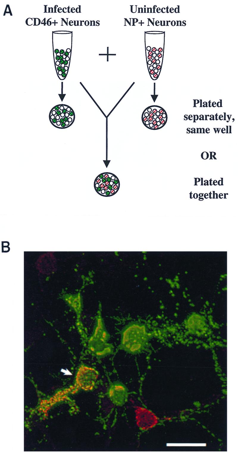
FIG. 6.

(A) Diagram of neuron coculture experiments, showing infected CD46+ cells (green with checkerboard, indicating staining for MV antigen with fluorescein isothiocyanate) and NP+ cells (red stripe, indicating staining for NP with rhodamine red-X) cultured together or on separate coverslips. The NP transgene was detected only on approximately 50% of transgenic neurons. (B) Mixture of CD46+ and NP+ cells on the same coverslip, 3 d.p.i., fluorescently immunostained for MV antigen with fluorescein isothiocyanate (green) and for NP protein with rhodamine red-X (red). Colocalization, shown by yellow (arrow), indicates MV infection of NP+ cells. Bar = 25 μm.
