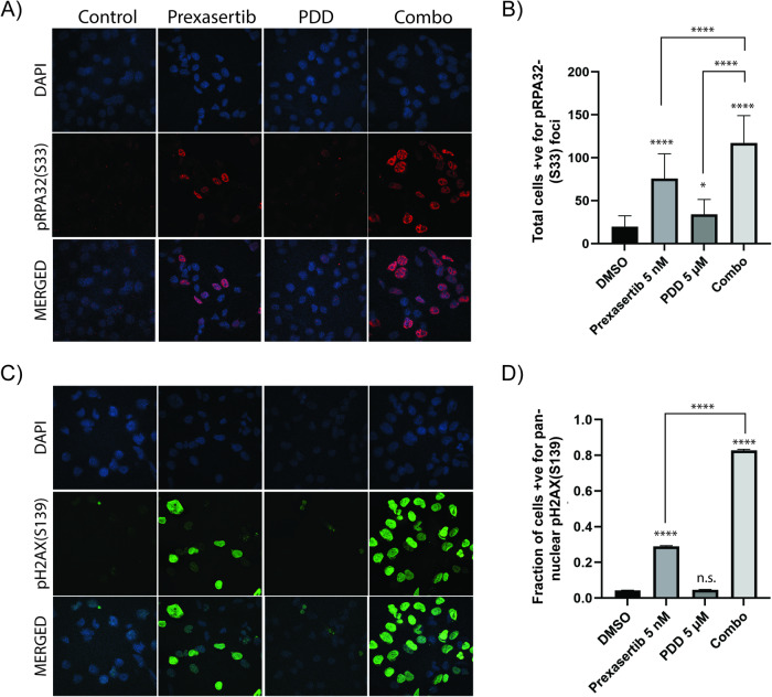
Fig. 4. Combination of prexasertib and PDD treatment causes replication stress and DNA damage.
A Immunofluorescence study showing pRPA32(S33) foci in SKOV3 cells treated with DMSO, 5 nM prexasertib, 5 µM PDD and their combination. B Histogram representation of total cells positive for pRPA32(S33) foci in SKOV3 cells treated with DMSO, 5 nM prexasertib, 5 µM PDD and their combination. A total of 50 cells from each of the three different experiments were counted for our histogram. C Immunofluorescence study showing pH2AX(S139) foci in SKOV3 cells treated with DMSO, 5 nM prexasertib, 5 µM PDD and their combination. D Histogram representation of percentage of cells with total pan-nuclear pH2AX(S139) staining in SKOV3 cells treated with DMSO, 5 nM prexasertib, 5 µM PDD and their combination. A total of 50 cells from each of the three different experiments were counted for our histogram. Error bars represent the mean ± standard deviation in case of pRPA32(S33) foci and mean ± standard error of mean (SEM) in case of pH2AX(S139). One-way ANOVA using Tukey’s multiple comparison tests were performed to analyze the statistical significance. n.s. not significant; *p < 0.05; ****p < 0.0001.

