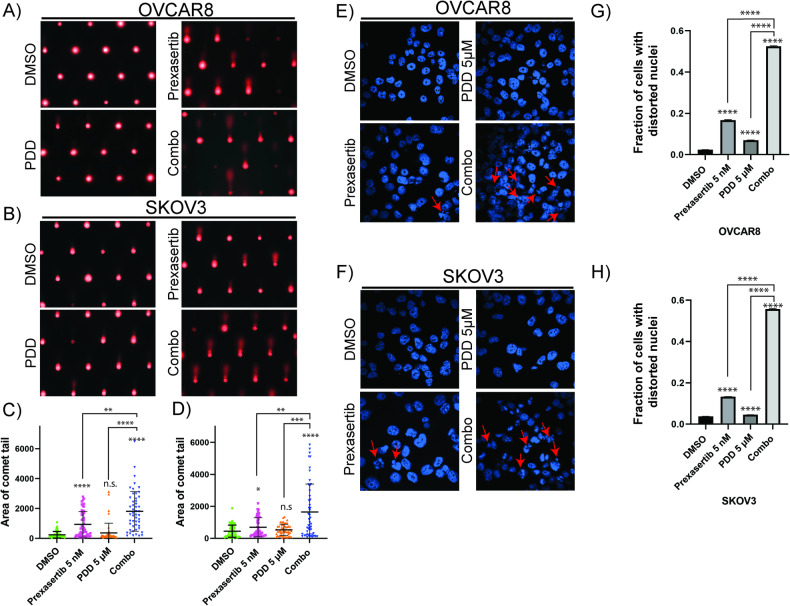
Fig. 5. PDD sensitizes prexasertib-induced DNA damage and increases nuclear distortion.
A, B Comet assay representative images treated with DMSO, 5 nM prexasertib, 5 µM PDD and their combination for 24 h in OVCAR8 and SKOV3, respectively. C, D Analysis of comet tail area in more than 50 cells from three independent experiments with their standard deviation as the error bars in OVCAR8 and SKOV3, respectively. E, F Distorted nuclei representative images in DAPI-stained nucleus of OVCAR8 and SKOV3, respectively treated with DMSO, 5 nM prexasertib, 5 µM PDD and their combination for 24 h. G, H Percentage of cells with distorted nuclei analyzed more than 200 cells from three different experiments with their standard error of mean as the error bars. One-way ANOVA using Dunnett’s T3 multiple comparison test for comet assay and Tukey’s multiple comparison tests for nuclear distortion assay were performed to analyze the statistical significance. n.s. not significant; *p < 0.05; **p < 0.01; ***p < 0.001; ****p < 0.0001.

