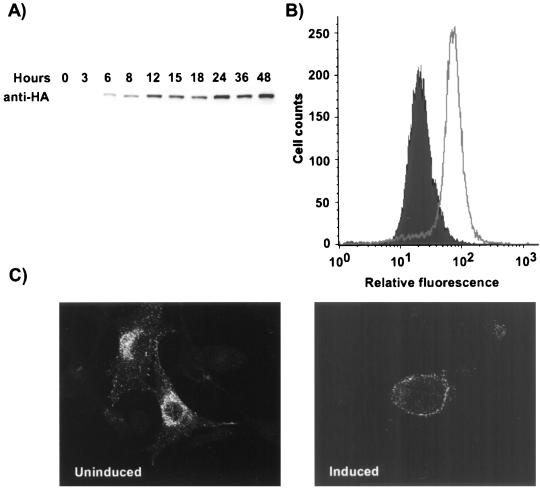
FIG. 3.
Characterization of Mv1-K44A cells. (A) Western blot analysis of dynamin K44A expression in Mv1-K44A cells at various times after tetracycline removal. The dynamin K44A was detected with MAb 12CA5 against the N-terminally fused HA epitope. (B) Expression of dynamin K44A in Mv1-K44A cells. Cells grown with or without tetracycline for 48 h were harvested for flow cytometry. The HA-tagged dynamin was detected by indirect immunofluorescent staining with MAb 12CA5, and 20,000 cells were analyzed by flow cytometry. (C) Uptake of HuTfn by induced and uninduced Mv1-K44A cells. Mv1-K44A cells transfected with the HuTfnR gene were grown in the presence or absence of tetracycline for 48 h on coverslips. Texas red-labeled HuTfn was incubated with the cells for 10 min at 37°C prior to fixation and examination by fluorescence microscopy.

