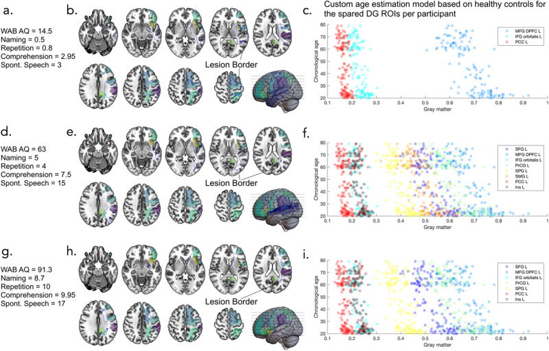
Fig. 6. Brain age estimations for domain-general regions.
Brain age estimation of domain-general regions for 3 example participants to highlight the use of different ROIs for each individual brain age estimation based on which ROIs were spared by the lesion. show the first participant, where (a) gives an outline of their behavioral scores, (b) shows an outline of their lesion (blue line) alongside the left domain general ROIs, and (c) shows the custom age estimation model based on healthy controls for the spared domain-general ROIs for this participant (note they are different for each participant based on which ROIs are lesioned). Similarly, (d, g) show behavioral scores for 2 other participants, (e, h) show brain maps of the overlap between their lesions and the domain general ROIs, and (f, i) show their custom age estimation models. Note that figures are in neurological orientation so the left (lesioned) hemisphere is shown on the right. WAB AQ Western Aphasia Battery Aphasia Quotient, Spont. Speech Spontaneous Speech, DG domain general, ROIs regions of interest, MFG DPFC L middle frontal gyrus dorsal prefrontal cortex left, IFG orbitalis L inferior frontal gyrus orbitalis left, PCC L posterior cingulate gyrus left, SFG L superior frontal gyrus left, PrCG L precentral gyrus left, SMG L supramarginal gyrus left, Ins L insular left.

