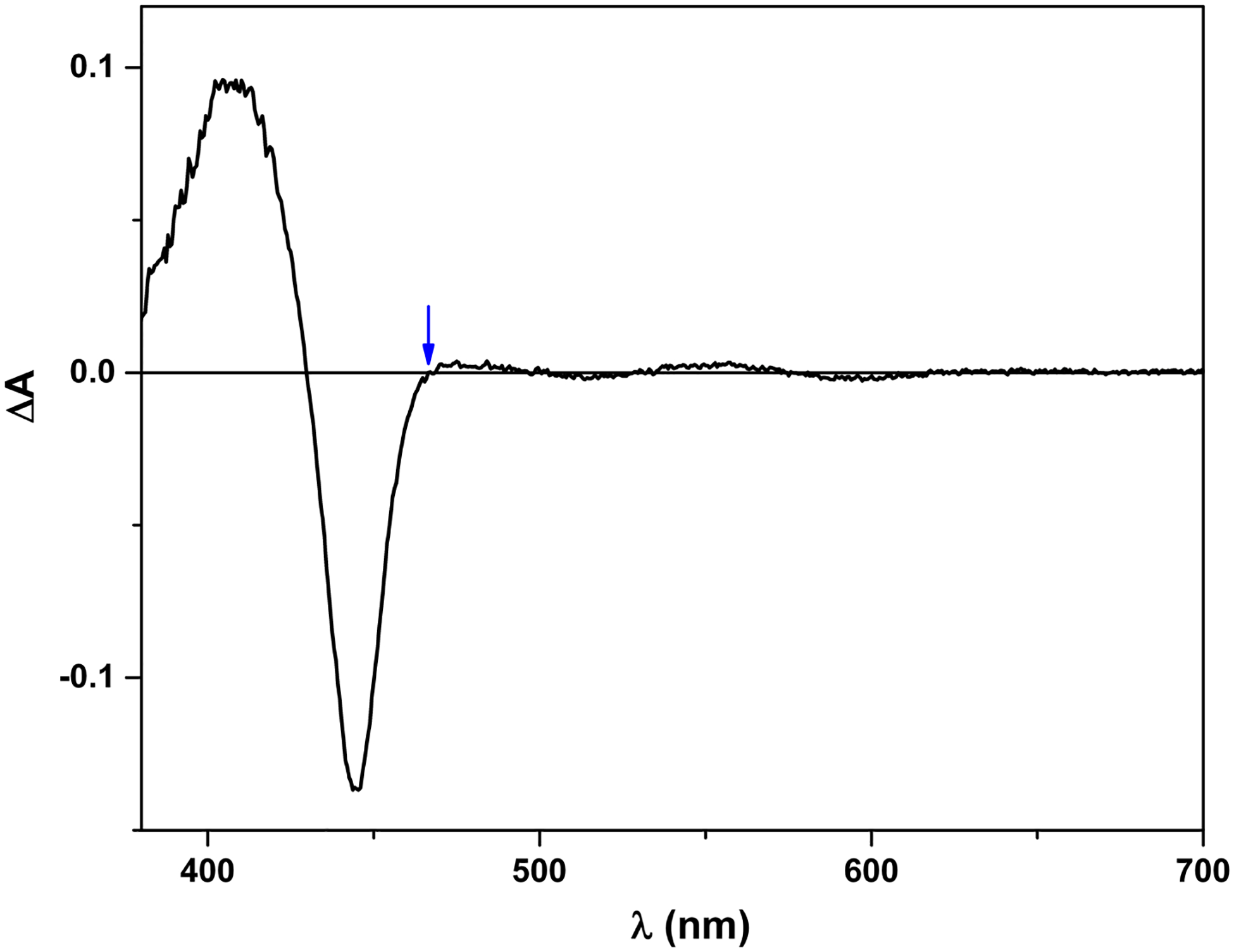
Figure 5.

Laser-induced difference spectrum of iNOSoxy. The protein was first reduced to Fe2+-CO form, and then excited by 450 nm laser. The difference spectrum was collected on an Edinburgh L900 laser flash photolysis instrument in spectral mode using an ANDOR iCCD camera. Note that 465 nm is an isosbestic point in the spectrum (marked by an arrow). In other words, the CO rebinding process does not contribute to the absorbance change at 465 nm, which gives a (narrow) window to observe re-oxidation of Fe2+ (due to the FMN-heme IET).
