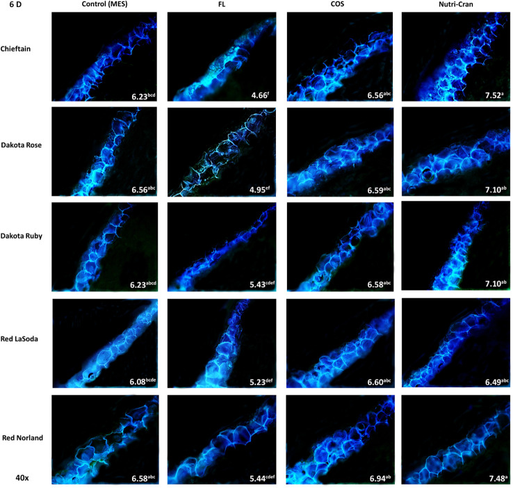
Figure 3.
Formation of SPP at the wounded surface of potato tuber tissues (cvs. Chieftain, Dakota Rose, Dakota Ruby, Red LaSoda, and Red Norland). Picture depicts the variations in the SPP formation (40x magnification) with different treatments (Control, fluridone-FL, chitosan oligosaccharide-COS, and cranberry pomace residue- Nutri-Cran) at 6 d after wounding. At 6 d (B), SPP accumulation completed the first cell layer in control sample and spread to second cell layers with elicitor treatments. In Chieftain, Dakota Ruby, and Dakota Rose, accumulation of SPP was not even complete at first cell layer with FL treatment, indicating a significant inhibition. Different lowercase letters represent statistically significant differences between cultivar × treatment interaction based on Tukey’s HSD test at 95% confidence level.

