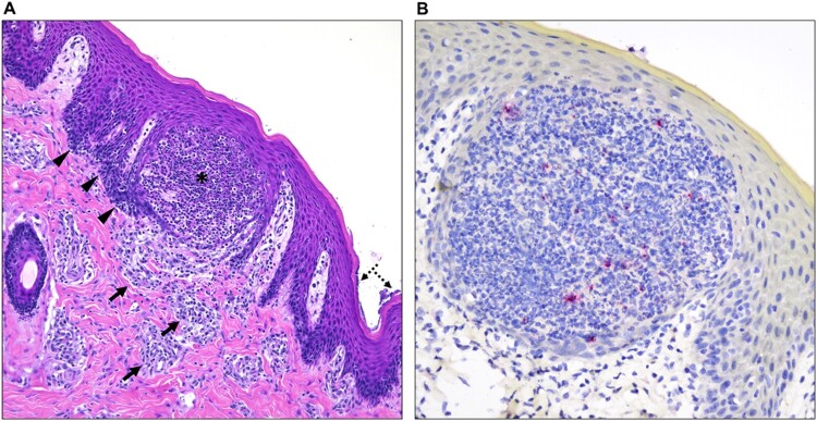
Figure 2.
Histological alterations in the skin and immunohistochemical detection of MPXV antigen in one of the infected pigs. (A) Histologic alterations are characterized by epidermal pustule formation (*), hyperplasia of the adjacent epidermis (arrowheads) and superficial perivascular lymphohistiocytic and eosinophilic dermatitis (arrows). The corneal layer of the epidermis was occasionally colonized by coccoid bacteria (dashed arrows). (B) Within this single pustule, sporadic degenerate inflammatory cells contain minimal intracytoplasmic viral antigen (red). No viral antigen was detected within keratinocytes or other cell types. H&E, 200X total magnification; Fast Red, 400X total magnification.

