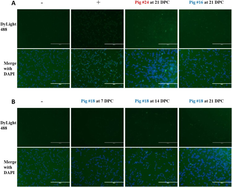
Figure 6.
Detection of MPXV-specific antibodies using Protein A. Vero E6 cells were infected with MPXV at a MOI of 1 and incubated for 48 hours before fixation with 80% acetone. Pig sera were diluted in PBS with 1% BSA and added to fixed wells. Biotinylated Protein A conjugated to streptavidin-DyLight488 was used to stain wells, and wells were visualized at 10x magnification using an EVOS fluorescence microscope. Scale bars represent 400 µm. (A) Representative images of stained cells. Naïve serum collected at -1 DPC was used as a negative control, and a rabbit anti-vaccinia polyclonal antibody was used as a positive control. (B) Images from sentinel pig #18, collected at each indicated timepoint.

