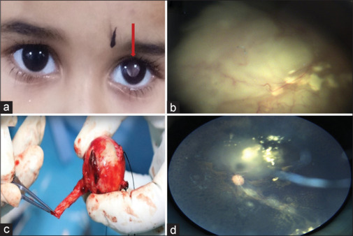
Figure 1.

(a) Clinical photograph illustrating white reflex in the left eye (red arrow). (b) Fundus image showing group E retinoblastoma with a large endophytic tumor and diffuse subretinal fluid. (c) Clinical image showing enucleated eyeball with 15-mm optic nerve. (d) Fundus image showing regressed tumor (Type 3) following three sessions of intra-arterial chemotherapy
