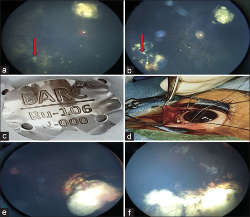
Figure 2.

(a) Fundus image showing active vitreous seeds (red arrow). (b) Fundus image showing regressed and calcified vitreous seeds following three sessions of intravitreal melphalan. (c) Clinical photograph showing indigenous ruthenium plaque. (d) Intraoperative image showing the ruthenium plaque over the tumor. (e) Fundus photograph showing recurrence of tumor in the inferotemporal quadrant over the edge of chorioretinal scar of cryotherapy with feeder vessel at the periphery with intrinsic vascularity. (f) Fundus photograph showing completely regressed tumor after 1 month
