FIG. 5.
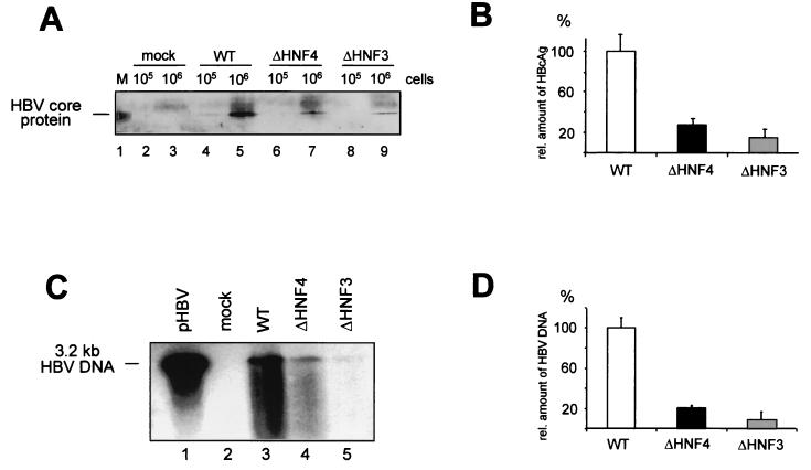
The replication competence of enhancer I mutants is impaired. HuH-7 cells were transfected with the empty vector control (pBS; mock), the wt, and the enhancer I ΔHNF3 and ΔHNF4 HBV replication-competent constructs. (A and B) Quantification of intracellular core protein expression. (A) Core protein was precipitated by monoclonal mouse anti-HBcAg-specific antibodies. The precipitated protein was run on SDS-PAGE and transferred to nylon membranes. Filters were probed with polyclonal rabbit anti-HBcAg antiserum. Either 105 or 106 cells were used in the experiments as indicated. In lane 1, recombinant core protein was used. (B) The relative amounts of HBcAg were determined by Fuji phosphoimager or densitometric evaluation. The amount found with the wt HBV construct was set to 100%. The error bars indicate standard deviations. (C and D) The amount of newly synthesized progeny DNA was determined from cytoplasmic HBV core particles by using anti-HBc antibodies for immunoprecipitation from 106 cells. (C) HBV DNA was separated on a 1% alkaline agarose gel and visualized by Southern blot analysis as described previously (4). A 3.2-kb linear HBV DNA was used as a reference (lane 1). (D) The concentration of progeny HBV DNA was quantified; wt values were set to 100%, and the amounts of the HNF3 and HNF4 mutants were calculated. The error bars indicate standard deviations.

