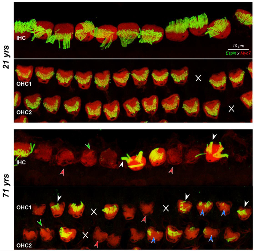
Figure 3:

Confocal images showing IHCs and the first two rows of OHCs with their stereocilia bundles, at the 5.6 kHz region from the 21 yr old and the 71 yr old subject. White X’s indicate missing OHCs. Red arrowheads denote hair cells with no stereocilia, blue arrowheads indicate foci of missing stereocilia, and white arrowheads point to fusion bundles. Green arrowheads point to stereocilia bundles visible in the myosin channel that did not stain with espin. Scale bar at the top applies to all panels.
